The Ophthalmoscope, also known as Funduscope, is a device that brought an explosive change in the treatment of eye diseases. It is used to find eye diseases such as Glaucoma. Before the device, the physicians had been relying primarily on magnifying glasses to study the eyes. It was more or less unreliable, as the doctors could only see the outer side of the eye and it was difficult to do the diagnosis. Eye is a very complex organ containing many microscopic blood vessels, lens and retina. And, unlike many of the other organs, eye couldn’t be cut open to examine either. The invention of Ophthalmoscope was therefore a major turning point in the field.
History of Opthalmoscope
The idea behind Ophthalmoscope was first suggested by English physician Dr. William Cumming in 1846 in an essay. According to some historians, the first Ophthalmoscope was the contribution of English mathematician Charles Babbage in 1847. It is claimed that Babbage, also known as the father of computer, gave his device to a doctor to check but it didn’t see the light ever since. But the invention of a practical Ophthalmoscope is widely attributed to a German physician named Hermann von Helmholtz. He developed his own version of the Ophthalmoscope in 1851, without any knowledge of Babbage’s achievement.
Helmholtz reached his invention after many experiments on magnifying glasses. His device used a mirror to direct a light beam to eye and using lenses, a much larger image of the eye could be obtained. The apparatus helped one to see the small veins and other minute parts of the eye. Later, Swedish ophthalmologist Allvar Gull-strand developed an improved version of the device. In 1915, Francis A. Welch and William Noah Allyn invented the world’s first hand-held direct illuminating ophthalmoscope which is often regarded as the model for the current Ophthalmoscopes. They established a company, Welch Allyn, which is one of the largest manufacturers of the device now.
Structure
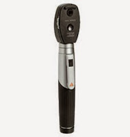
An Ophthalmoscope helps a physician to take a detailed observation of the interior parts of the eye and find any ailments and signs of diseases. The basic structure of Ophthalmoscope is almost common in all versions. Here are the features present in an opthalmoscope:
Aperture
Aperture enables the devices to be used in different ways by modifying the amount of light that passes through the lens.
Rheostatic Switch
It is a control switch which powers the light source. Practitioners can manually adjust the brightness of light through this switch.
Lens Selection Wheel
There are multiple viewing lenses varying by device type.
Focusing Wheel
As the name suggests, the wheel is used to focus on the eye of the patient.
Brow Rest
This rest allows the examiner to wear glasses when using the ophthalmoscope.
How to use an Ophthalmoscope
Before using the ophthalmoscope, you need to clear the area around you and have plenty of lighting options as well as enough room for both examiner and patient to move around. Dim the lights of the room as it helps dilate the pupil of the eye. Practitioners also put dilating drops for maximum viewing capabilities. Using drops is optional.
Next, ask the patient to focus on one particular object as you set the device to maximum light. Begin by performing red reflex test to check the reflection of light from the retina. Improper or no reflection means there might be a disorder in the retina. As the ophthalmoscope is six inches from the patient, look into the patient’s eye. Adjust the focusing wheel until your view is crisp. Follow the arteries, examine the vascular arcades and evaluate the macula of the eye. Clean all the tools properly after you are done with the examination.
Types
There are two types of ophthalmoscopes, direct (traditional) and PanOptic (indirect) ophtlamoscope. Direct Ophthalmoscope allows you to check the back of the eye to observe the retina, optic nerve, vasculature and vitreous humor. An exam like this produces an upright image that is approximately 15 times magnified. Indirect opthalmoscope produces an inverted image of your retina which is approximately 2x or 5x magnified.
Benefit of Opthalmoscope
With their bright lights and refined lenses, ophthalmoscopes can provide a clear view of the fundus of the eye. This view can reveal much about a person’s health. At present, Ophthalmoscope is used to determine even high Blood pressure and Diabetes. The practice of using the device to determine the eye diseases is called Ophthalmoscopy or Funduscopy. Ophthalmoscopy can bring into light the non-urgent conditions of the eye like a cataract. It can also detect foreign bodies in the cornea and pupil distortions.
Related posts: